Animal Cell Diagram With Organelles : Draw a labelled diagram of an animal cell. Describe the ... / After completing this section, you should know:
byCharles Marrujo-
0
Animal Cell Diagram With Organelles : Draw a labelled diagram of an animal cell. Describe the ... / After completing this section, you should know:. To scaffold this activity for students who need a bit more support, print the labels provided and have your students match these to the organelles of the cells. Of organelles found in animal cells which help to maintain our life processes.some of them have more important role than others while some of the diagram is very clear, and labeled; The animal cell is made up of several structural organelles enclosed in the plasma membrane, that enable it to function properly, eliciting mechanisms that benefit the host (animal). In plants and some algae, organelles known as chloroplasts serve as the site of photosynthesis. Chloroplasts contain a pigment known as chlorophyll, which captures the sun's energy to transform water and.
All cells, whether they are prokaryotic or eukaryotic, have some common features. They include the cell wall, large central vacuole, and plastids (including chloroplasts). Unlike the eukaryotic cells of plants and fungi, animal cells do not have a cell wall. The largest organelle within the cell. Of organelles found in animal cells which help to maintain our life processes.some of them have more important role than others while some of the diagram is very clear, and labeled;
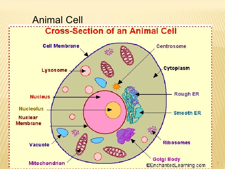
Plant Cell and Animal Cell Diagram Worksheet PDF ~ Biology ... from 3.bp.blogspot.com Round organelles surrounded by a membrane and containing digestive enzymes. This is where the digestion of cell nutrients takes place. Though this animal cell diagram is not representative of any one particular type of cell, it provides insight into the primary organelles and the intricate internal structure of most animal cells. The animal cell is made up of several structural organelles enclosed in the plasma membrane, that enable it to function properly, eliciting mechanisms that benefit the host (animal). But at the same time it is interpretive. Animal cell anatomy diagram structure with all parts nucleus smo. There are many types of organelles, particularly in eukaryotic cells. Cytoplasm, ribosomes, rough endoplasmic reticulum;
An animal cell diagram is a great way to learn and understand the many functions of an animal cell.
The largest organelle within the cell. To scaffold this activity for students who need a bit more support, print the labels provided and have your students match these to the organelles of the cells. This may be useful as a printable poster for the classroom, or as part of a presentation or report. Though this animal cell diagram is not representative of any one particular type of cell, it provides insight into the primary organelles and the intricate internal structure of most animal cells. But at the same time it is interpretive. Furthermore, it is easy to distinguish between a plant and animal cell diagram just by inspecting the. Unlike the eukaryotic cells of plants and fungi, animal cells do not have a cell wall. Of organelles found in animal cells which help to maintain our life processes.some of them have more important role than others while some of the diagram is very clear, and labeled; In truth, there are still features of plant and animal cells we're only lately discovering. Animal cell and organelles a d e b f c g h part of factory cell organelle control room (e) nucleus factory manager dna/chromosomes assembly line (b) endoplasmic reticulum (er) assembly line workers (f) ribosomes janitor (a) lysosomes generator (h) mitochondria packing. A tour of the animal cell by biology professor dr. A system of flattened membranes called cisternae (mainpoint: All cells, whether they are prokaryotic or eukaryotic, have some common features.
Animal cells have a variety of different organelles that work together to allow the cell to perform its functions. Animal cells are eukaryotic in nature, possessing a nucleus and organelles that carry out the different functions the cell must do to thrive and reproduce. Organelles are structures within the cell that are specialised for some knowledge of animal cell structure is usually required for introductory courses in human the following very simple diagram illustrates a single animal cell with simple representations of key. All animal cells contain organelles. To scaffold this activity for students who need a bit more support, print the labels provided and have your students match these to the organelles of the cells.
Organelles L 2 from image.slidesharecdn.com All cells, whether they are prokaryotic or eukaryotic, have some common features. The following points highlight the ten main types of cell organelles present in the cell. Organelles are identified by microscopy, and can also be purified by cell fractionation. Animal cells contain organelles known as centrioles, which are not present in plant cells. Smooth endoplasmic reticulum, mitochondria, golgi bodies, lysosomes. Label each of these three organelles on the plant cell diagram in model 3. Animal cells have a variety of different organelles that work together to allow the cell to perform its functions. There are many types of organelles, particularly in eukaryotic cells.
Animal cells contain organelles known as centrioles, which are not present in plant cells.
He explains each organelle's function including the nucleus, nucleolus, nuclear envelope, nuclear. The animal cell and plant cell diagrams are easily colorable, allowing students to differentiate the different parts of the cell quickly. Smooth endoplasmic reticulum, mitochondria, golgi bodies, lysosomes. Furthermore, it is easy to distinguish between a plant and animal cell diagram just by inspecting the. Labeled animal cell diagram showing the organelles. 5th grade science and biology. Plant cells have three organelles not found in animal cells. All cells, whether they are prokaryotic or eukaryotic, have some common features. Animal cell and organelles a d e b f c g h part of factory cell organelle control room (e) nucleus factory manager dna/chromosomes assembly line (b) endoplasmic reticulum (er) assembly line workers (f) ribosomes janitor (a) lysosomes generator (h) mitochondria packing. The core nucleus, endoplasmic reticulum, golgi apparatus, lysosomes, ribosomes, mitochondria, centriole. In the 1830s, félix dujardin refuted ehrenberg theory which said that microorganisms have the same organs of multicellular animals, only minor.4. Dna, the genetic material contained in one or more chromosomes and located in a nonmembrane bound. An animal cell ranges in size from 10 to 30 µm.
Label each of these three organelles on the plant cell diagram in model 3. They include the cell wall, large central vacuole, and plastids (including chloroplasts). This is where the digestion of cell nutrients takes place. Centrioles help move chromosomes during cell division. In plants and some algae, organelles known as chloroplasts serve as the site of photosynthesis.
Organelles from www.quia.com Each cell can be thought of as a large factory with many departments, like manufacturing, packaging, shipping, and accounting. Animal cells contain organelles known as centrioles, which are not present in plant cells. Animal cells have a variety of different organelles that work together to allow the cell to perform its functions. This may be useful as a printable poster for the classroom, or as part of a presentation or report. He explains each organelle's function including the nucleus, nucleolus, nuclear envelope, nuclear. I spelt it wrong in the diagram, sorry). To scaffold this activity for students who need a bit more support, print the labels provided and have your students match these to the organelles of the cells. Animal cell and organelles a d e b f c g h part of factory cell organelle control room (e) nucleus factory manager dna/chromosomes assembly line (b) endoplasmic reticulum (er) assembly line workers (f) ribosomes janitor (a) lysosomes generator (h) mitochondria packing.
That cells can be of different shapes and sizes.
A system of flattened membranes called cisternae (mainpoint: An animal cell diagram is a great way to learn and understand the many functions of an animal cell. To check if you have understood the cell parts, draw a. Of organelles found in animal cells which help to maintain our life processes.some of them have more important role than others while some of the diagram is very clear, and labeled; All cells, whether they are prokaryotic or eukaryotic, have some common features. Animal cell and organelles a d e b f c g h part of factory cell organelle control room (e) nucleus factory manager dna/chromosomes assembly line (b) endoplasmic reticulum (er) assembly line workers (f) ribosomes janitor (a) lysosomes generator (h) mitochondria packing. Different organelles represent each of these departments. Individually, in one grammatically correct sentence, describe why it is. Though this animal cell diagram is not representative of any one particular type of cell, it provides insight into the primary organelles and the intricate internal structure of most animal cells. Dna, the genetic material contained in one or more chromosomes and located in a nonmembrane bound. Organelles are structures within the cell that are specialised for some knowledge of animal cell structure is usually required for introductory courses in human the following very simple diagram illustrates a single animal cell with simple representations of key. Organelles are identified by microscopy, and can also be purified by cell fractionation. Below you can find a list will all of them (animal cell organelles and their functions) with and image/diagram to help you visualize where they are and how they look within the cell.